Research
We study the neural mechanisms underlying visual perception in healthy and clinical populations. We focus on motion and depth perception, as they reveal the transformations of sensory input into perceptual state as neural signals travel through the brain.
Our research helps uncover the function and underlying architecture of the brain’s visual system, provides us with a model by which to understand sensory processing in general, and contributes to the treatment of perceptual impairments such as amblyopia (lazy eye). We use complementary behavioral, neuroimaging and computational approaches.
The Laboratory is affiliated with the Psychology Program and Brain Imaging Facility at New York University Abu Dhabi as well as the Department of Psychology and Center for Neural Science at New York University.

Altered White Matter in Early Visual Pathways of Humans with Amblyopia
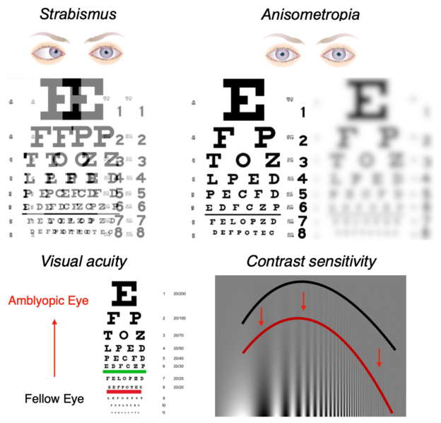
Amblyopia (better known as lazy eye) is a disorder where vision is reduced, without a clear deficit in the eyes themselves. It occurs when the images from our two eyes are poorly correlated during development. Over time the brain tends to suppress the image from the ‘lazy’ eye, in favor of the ‘good’ eye.
Such suppression leads to abnormal visual experience, and as such amblyopia is of interest not only as a clinical disorder, but also as a human model of the interaction between sensory input and brain development.
In our research, we asked what the neuroanatomical consequences of visual suppression are. We show that amblyopia leads to structural changes in early visual pathways, connecting the thalamus to striate and extrastriate visual cortex. This is most likely due to changes in cortico-thalmic feedback projections. Our work gives us a clearer picture of visual development and benefits the diagnosis and treatment of visual disorders such as amblyopia.
See our paper in Vision Research for more information.

Motion Processing with Two Eyes in Three Dimensions
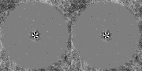
Two eyes, three dimensions The movement of an object toward or away from the head is perhaps the most critical piece of information an organism can extract from its environment. Such 3D motion produces horizontally opposite motions on the two retinae.
Canonical conceptions of primate visual processing assert that neurons early in the visual system combine monocular inputs and extract 1D (component) motions; later stages then extract 2D pattern motion from the output of the earlier stage. We found, however, that 3D motion perception is in fact affected by the comparison of opposite 2D pattern motions from each eye individually.
These results imply the existence of eye-of-origin information in later stages of motion processing and therefore motivate the incorporation of such eye-specific pattern-motion signals in models of motion processing and binocular integration.
A paper on our findings has been published in the Journal of Vision.

Neural Circuits Underlying the Perception of 3D Motion
The macaque middle temporal area (MT) and the human MT complex (MT+) have well-established sensitivity to both 2D motion and position in depth. Yet evidence for sensitivity to 3D motion has remained elusive.
We showed that human MT+ encodes two binocular cues to 3D motion, one based on estimating changing disparities over time, and the other based on interocular comparisons of retinal velocities.
By varying orientation, spatiotemporal characteristics, and binocular properties of moving displays, we distinguished these 3D motion signals from their constituents, instantaneous binocular disparity and monocular retinal motion. Furthermore, an adaptation protocol confirmed MT+ selectivity to the direction of 3D motion.
These results demonstrate that critical binocular signals for 3D motion processing are present in MT+, revealing an important and previously overlooked role for this well-studied brain area.
A paper on our findings has been published in Nature Neuroscience. You can also view animations of the stimuli.

